
Despite the fact that the convenience of planning the day of birth has serious downsides, as many as 40% or more of mothers today choose…
A blog about everything related to pregnancy and childbirth
Despite the fact that the convenience of planning the day of birth has serious downsides, as many as 40% or more of mothers today choose…

There are many ways to write a birth plan. Here are some basic guidelines about what to do as well as what not to do…

For many years, laboring women were encouraged to utilize a type of pushing during childbirth known as “Valsalva.” This technique involved holding one’s breath for…

Walking epidurals have become all the rage when it comes to the most popular trends for childbirth today. One of the biggest complaints in the…
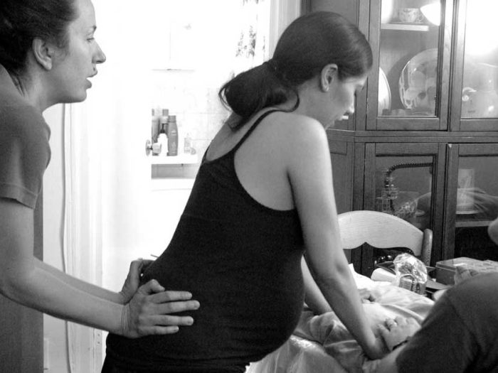
Medical Benefits of Hiring a Doula Numerous studies in the last 25 years have clearly demonstrated the value of a birth doula during the process…

Being a new mum is one of the most exciting journeys you can experience, but it’s no secret that its financial challenges can be overwhelming.…

It is incredible how less observant women won’t know they are pregnant until months later when they are almost past the first trimester. Those with…

The numbers of labor inductions are increasing today. It seems that there are a host of reasons that a care provider may recommend an induction,…

Since the earliest beginnings of childbirth education, educators have emphasized using breathing exercises as a complement to relaxation during labor. Here are even more reasons to…

By the time you reach the halfway point of your pregnancy, you will start to feel your tummy tightening up for a minute or so…